Histology of the Feline Eye: My Favourite Tissue
Main Function: The cat eye is the main visual sensory organ of the cat. The feline eye's function is to detect light and convert it to chemical signals that can be interpreted by the feline brain. Cat eye's contain most characteristics associated with human eyes except for the well-known reflective layer located behind the retina called the tapetum lucidum (Wrycha, 2004).
Main Anatomy of the Feline Eye:

Figure 1: Basic anatomy of the feline eye globe. (Wrycha, 2004)
Anterior Chamber: This chamber is covered by the cornea. The cornea receives light signals and directs them towards the lens. The cornea serves as a window to the eye and has great refracting power. The stroma makes up the majority of the cornea and is mainly composed of lamellae with sparse collagen fibres. The Anterior Chamber is filled with aqueous fluid (Wrycha, 2004).

Figure 1: Feline cornea showing the outer epithelium (stratified squamous), stroma and the endothelium (simple cuboidal) (Wrycha, 2004).
Posterior Chamber: This chamber contains the lens, and it is separated from the anterior chamber via the iris'. The iris and retina are very sensitive to light and may close around the pupil in a "slit-like" manner when exposed to bright light intensities (Wrycha, 2004).
The Lens: This convex structure serves to receive refracted light from the cornea and to refract and focus the light towards the retina.
The Iris: This structure accounts for the vibrant colours observed in the variety of feline eyes (as well as human eyes). The iris also contains a layer of muscle so that it can contract and dilate over the pupil in response to bright and low light intensities.

Figure 2: Histological section of a Feline Iris. "M" designates the melanocytes that give the iris colour while "U" designates the dilator muscle of the iris (Wrycha, 2004).
The Retina: This structure lines the inside of the feline eye and is the site of the conversion of light signals to chemical signals. Chemical signals are then transferred into electrical signals via nerve impulses sent from the Optic Nerve. The important cells within the Retina are the "Rods" and "Cones". Rods specialize in detecting light in low light intensities while cones specialize in detecting light in bright light intensities. Cones are also responsible for colour vision.

Figure 3: Histological section of the Feline Retina. "A" and "B" designate the thick layer of rod and cone cells. "T" designates the Tapetum (Wrycha, 2004).
INTERESTING FACT!
The layer of "rod" cells in cats are thicker than the rod cells in dogs. This extra thickness accounts for the reason why cats see much better in the dark than humans or dogs!
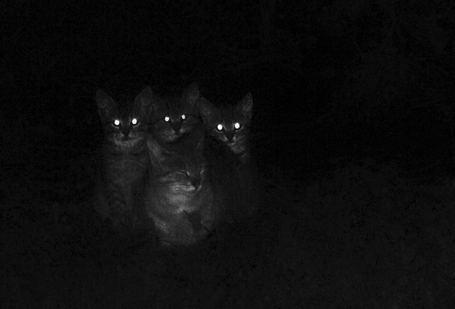
Figure 4: Cats being creepy in the dark. (Those are their tapetums shining!) (Rodo, 2010)
Sclera: The white area of the eye that contains collagen and elastic fibres. In cats, the sclera is not visible unless the eye-lid is pulled back (the iris takes up the visible area of the eye (unlike humans) (Sclera, 2012)
Pathology of the Feline Eye (The sad section)
This section will outline the most common problems and diseases that occur in the eye's of cats. Grab some tissues.
1. Conjunctivitis: This is the most common pathology observed in cats. It is similar to pink eye that we see in humans. It involves the swelling and irritation of the inner lining of the eye-lid or cornea. Conjunctivitis in cats is almost always indicative of another infection (i.e. herpes virus or chlamydophila). Without treatment, conjunctivitis can lead to serious vision problems. Most treatments entail the application of a topical, antibiotic cream to the eyes. (Carlson & Giffin, 2008)

Figure 5: Kitten showing conjunctivitis in the eyes. (From "M's" blog, 2011)
2. Cataracts: This problem is associated with an opacity on the lens of the cat's eye. The opacity prevents light from passing through the lens and ultimately renders the cat partially blind (Cataracts in Cats, 2011)

Figure 6: Cat showing cataract in one eye (Don's Blog, 2010).
3. Feline Glaucomas: Glaucomas are diseases that are characteristically neuro-degenerative and result in the degeneration of the retinal and optic nerves. This degeneration results in the lost of vision for the cat. Glaucomas typically arise through elevated levels of intra-ocular pressure. Therefore the most common treatment used it the administration of hypo-tensive drugs (McLellan & Miller, 2012).

Figure 7: Cat eye displaying typical features of glaucoma. (Ferreira, 2010)
References:
Cataracts in Cats (2011, October 22). Retrieved from:
http://www.pethealthnetwork.com/cat-health/cataracts-cats
Carlson, D., and J.M. Giffin. (2008). Conjunctivitis (Pinkeye) in Cats: Types, Symptoms, Causes and
Treatments. Retrieved from: http://pets.webmd.com/cats/conjunctivitis-pinkeye-cats-types-symptoms-causes-treatments?page=2
McLellan, G.J., and P.E. Miller. (2012). Feline Glaucoma- A Comprehensive View. Retrieved From:
Sclera (2012). Retrieved from: http://www.stlukeseye.com/anatomy/sclera.html
Wrycha (2004). Anatomy and Histology of the Canine and Feline Eye. Madison: University of Wisconsin
No comments:
Post a Comment